
It involves placing vitamin-A drops in the eye and exposing it to a certain wavelength of ultraviolet light. Curr Opin Ophthalmol 2010 21(4):255.FDA trials are currently underway for a procedure called corneal cross-linking. Treatment strategies for corneal ectasia. Complications of intrastromal corneal ring segment implantation using a femtosecond laser for channel creation: a survey of 850 eyes with keratoconus. Corneal collagen cross-linking with riboflavin and ultraviolet a irradiation for keratoconus: long-term results.

Modified intracorneal ring segment implantations (INTACS) for the management of moderate to advanced keratoconus: efficacy and complications. Collagen cross-linking with riboflavin and ultraviolet-A light in keratoconus: One-year results. Mechanisms of corneal tissue cross-linking in response to treatment with topical riboflavin and long-wavelength ultraviolet radiation (UVA) Invest Ophthalmol Vis Sci 2010 51:129–38.ġ6. McCall AS, Kraft S, Edelhauser HF, et al. Factors associated with corneal graft survival in the cornea donor study." JAMA Ophthalmol 2015 133(3):246–54.ġ5.

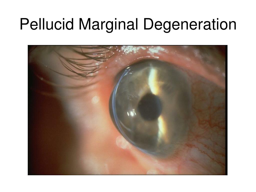
Scleral lenses in the management of keratoconus. A Guide to Scleral Lens Fitting, Version 2.0." Forest Grove, IL: 2015.ġ3. Barr JT, Wilson BS, Gordon MO, et al Estimation of the incidence and factors predictive of corneal scarring in the Collaborative Longitudinal Evaluation of Keratoconus (CLEK) Study. Pellucid marginal degeneration coexistent with cornea plana in one member of a family exhibiting a novel KERA mutation. Nagy M, Vigvary L: Etiology of the pellucid marginal degeneration of the cornea. Pellucid corneal marginal degeneration: A review. Jinabhai A, Radhakrishnan H, O’Donnell C. Do I see keratoconus or PMD? How can we differentiate between these two similar ectatic diseases on corneal topography? Rev Optometry 2005 142(4):91–92.Ĩ. Analysis of pseudoprogression after corneal cross‐linking in children with progressive keratoconus. Davidson AE, Hayes S, Hardcastle AJ, Tuft SJ. Collagen crosslinking with riboflavin and ultraviolet-A light in keratoconus: long-term results. Collaborative Longitudinal Evaluation of Keratoconus (CLEK) Study: methods and findings to date. The epidemiology and etiology of keratoconus. Gordon-Shaag A, Millodot M, and Shneor E. Jinabhai, Radhakrishnan H, and O’Donnell C. In addition, this paper shows an innovative way to utilize corneal topography to ensure proper fitting scleral lenses.ġ. This case demonstrates the usefulness of utilizing scleral gas permeable (GP) contacts over INTACS post CXL to provide optimal, stable vision, comfort and therapy. Regardless, the Federal Drug Administration (FDA) approval of KC treatment using CXL in April 2016 has shown excellent promise of improving the visual potential for patients with this progressive disease. More importantly, the cause of KC is still unknown. This means most of those found having KC have no known association with the disease. The Collaborative Longitudinal Evaluation of Keratoconus (CLEK) study found the genetic predisposition of KC to be at 13.5%, whereas other studies found it to be between 6–10%. KC is predominantly bilateral (90%) and according to recent studies demonstrates a prevalence of 2.3%. KC is a developmental anomaly in which a part of the cornea becomes thinner and bulges forward in a cone-shaped fashion as a result of non-inflammatory stromal thinning. Although there is no evidence of the prevalence and etiology of PMD, it may be postulated that PMD is poorly differentiated because this disease is often confused or used interchangeably with keratoconus (KC). It has been found more commonly in males and appears between the second and fifth decade of life showing no signs of ethnic predilection. PMD is a rare, progressive, degenerative corneal disease typically characterized as peripheral, bilateral, and inferior ectasia producing a crescent shape. This particular case demonstrates the effectiveness of new scleral lenses over a severe form of PMD that underwent corneal crosslinking (CXL) with riboflavin over intra-stromal corneal ring segments (INTACS). This case study provides an analysis of an individual affected by pellucid marginal degeneration (PMD).


 0 kommentar(er)
